Home » Without Label » Names Of Muscles : The Cranial Nerve Exam · Anatomy and Physiology - The action refers to the action of each muscle from the standard anatomical position.
Names Of Muscles : The Cranial Nerve Exam · Anatomy and Physiology - The action refers to the action of each muscle from the standard anatomical position.
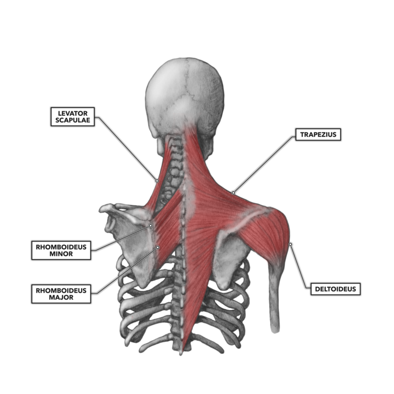
Names Of Muscles : The Cranial Nerve Exam · Anatomy and Physiology - The action refers to the action of each muscle from the standard anatomical position.. Included are several layered views of the back muscles, the doral muscles, subclavius muscles, rhomboideus major and minor muscles, deltoid muscles and many more. The following list contains a description of all of the possible functions of the muscles in the leg anatomy. A muscle of the medial thigh that originates on the pubis. There are three different types of muscles in the body: Related posts of body muscles with names head and neck muscle anatomy.
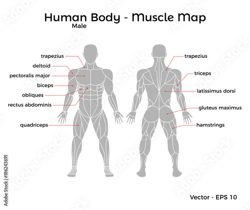
Many muscle names indicate the muscle's location. The muscles of the human body can be categorized into a number of groups which include muscles relating to the head and neck, muscles of the torso or trunk, muscles of the upper limbs, and muscles of the lower limbs. Three of them are located in the anterior compartment — the biceps brachii, brachialis, and coracobrachialis, while the forth is located in the posterior compartment — the triceps brachii). The deltoids, or delts, are known as the shoulder muscles. There are three different types of muscles in the body:
CrossFit | Shoulder Muscles, Part 2: Posterior Musculature from www.crossfit.com Use the find command to locate a specific muscle. Other muscle names can provide information as to how many origins a particular muscle has, such as the biceps brachii. A muscle of the medial thigh that originates on the pubis. Extends or adducts arm (swimming) flexes and rotates vertebral column. The human back extends from the buttocks to the posterior portion of the neck and shoulders. Each type differs in the muscles affected, the age of onset, and its rate of progression. The hamstring muscles in the back of the thigh, the quadriceps in the front and the adductor muscles on the inside. It is controlled by the obturator nerve.
Daniel nelson on january 1, 2019 2 comments 🔥!
Use the find command to locate a specific muscle. Included are several layered views of the back muscles, the doral muscles, subclavius muscles, rhomboideus major and minor muscles, deltoid muscles and many more. From there, the pectoralis major attaches to the collar bone (or clavicle) and converges on the upper arm bone (or humerus), just below the shoulder. The human back extends from the buttocks to the posterior portion of the neck and shoulders. Smooth muscle is under involuntary control and is found in the walls of blood vessels and of structures such as the urinary bladder, the intestines, and the stomach. Some muscles, like the tibialis anterior, are named after the part of the bone (the anterior portion of the tibia) that they are attached to. The cells of the muscles comprise protein filaments of actin and myosin that slide past one another, which produces contraction and changes both the length and the shape of the cell. It inserts onto the linea aspera of the femur. It is controlled by the obturator nerve. Sometimes, the way muscles interact with other muscles are incorporated into their names. Three of them are located in the anterior compartment — the biceps brachii, brachialis, and coracobrachialis, while the forth is located in the posterior compartment — the triceps brachii). By tightening and relaxing, the skeletal muscles create movement. These types of muscle tissue form the thick middle layer of the heart and are responsible for coordinated contractions,.
There are four muscles in you upper arm, which is delimited by your shoulder joint and your elbow joint. The thigh has three sets of strong muscles: Listed below are the 9 different types of muscular dystrophy. The most well known of the muscular dystrophies is duchenne muscular dystrophy (dmd), followed by becker muscular dystrophy (bmd). Included are several layered views of the back muscles, the doral muscles, subclavius muscles, rhomboideus major and minor muscles, deltoid muscles and many more.
Appendicular Muscles of the Pelvic Girdle and Lower Limbs ... from pressbooks-dev.oer.hawaii.edu Listed below are the 9 different types of muscular dystrophy. Broadly considered, human muscle—like the muscles of all vertebrates—is often divided into striated muscle (or skeletal muscle), smooth muscle, and cardiac muscle. The fixed end of a muscle is called the origin of the muscle. The pelvis at the bottom of the back and the shoulders at the top of the back give the back. From there, the pectoralis major attaches to the collar bone (or clavicle) and converges on the upper arm bone (or humerus), just below the shoulder. Latissimus dorsi is the name of the large muscles that run from under your arms, across your sides, and then across the middle of your back. Muscle name origin insertion function picture pectoralis major: Use the find command to locate a specific muscle.
I'll try to explain each movement by describing it in more universal terms instead of simply listing an exercise, which may be ambiguous or altogether unfamiliar to some.
Welcome to lumen's master muscle list see what happens when you have no muscles! Superficial and deep anterior muscles of upper body Muscle name origin insertion function picture pectoralis major: The thigh has three sets of strong muscles: Flexes and adducts (towards body) arm. Three of them are located in the anterior compartment — the biceps brachii, brachialis, and coracobrachialis, while the forth is located in the posterior compartment — the triceps brachii). They support bones, in this case, the vertebrae. Extends or adducts arm (swimming) flexes and rotates vertebral column. Broadly considered, human muscle—like the muscles of all vertebrates—is often divided into striated muscle (or skeletal muscle), smooth muscle, and cardiac muscle. The deltoids, or delts, are known as the shoulder muscles. Abductor digiti minimi (foot) abductor digiti minimi (hand). Lateral lip of bicipital groove of humerus and anterior lip of deltoid. A muscle of the medial thigh that originates on the pubis.
A muscle of the medial thigh that originates on the pubis. An involuntary, striated muscle type found in the heart. Abductor digiti minimi (foot) abductor digiti minimi (hand). Bodybuilders refer to these as their lats. so again, quick review, when it comes to size, the key identifiers are maximus or magnus, minimus, longus, brevis, and latissimus. Daniel nelson on january 1, 2019 2 comments 🔥!
Male Human Body Muscle map, with major muscle names, front ... from as1.ftcdn.net List of all leg muscles' functions. Latissimus dorsi is the name of the large muscles that run from under your arms, across your sides, and then across the middle of your back. Muscle is a soft tissue found in both animals and humans. Lateral lip of bicipital groove of humerus and anterior lip of deltoid. List of major anterior muscles major muscles on the front of the body adductor longus. Flexes and adducts (towards body) arm. Bodybuilders refer to these as their lats. so again, quick review, when it comes to size, the key identifiers are maximus or magnus, minimus, longus, brevis, and latissimus. Some muscle names indicate the number of muscles in a group.
The deltoid muscle consists of 3 parts:
This project was created with explain everything™ interactive whiteboard for ipad. Lateral lip of bicipital groove of humerus and anterior lip of deltoid. Listed below are the 9 different types of muscular dystrophy. Bodybuilders refer to these as their lats. so again, quick review, when it comes to size, the key identifiers are maximus or magnus, minimus, longus, brevis, and latissimus. It covers much of the front upper chest, beginning at the breastbone (or sternum) including the second to the sixth ribs. Daniel nelson on january 1, 2019 2 comments 🔥! Sometimes, the way muscles interact with other muscles are incorporated into their names. Some types are named for the affected muscles, including the. This anatomy chart is a great example of beauty and function in one, as it is pleasing to look at and is very educational… muscles of the shoulder, the arm and the forearm lateral view Head and neck muscle anatomy 12 photos of the head and neck muscle anatomy head and neck. The back muscles are skeletal muscles. The cells of the muscles comprise protein filaments of actin and myosin that slide past one another, which produces contraction and changes both the length and the shape of the cell. Anterior deltoid, medial deltoid, and posterior deltoid.